The Timepix detectors are hybrid active pixel detectors, which were developed within the Medipix collaboration at CERN (http://medipix.web.cern.ch/medipix) as the successors of the Medipix detectors.
The main part of a Timepix detector is the active sensor layer, which is segmented into a square matrix of 256 x 256 pixels with a pixel pitch of 55 um, thus covering an area of 1.4 x 1.4 cm2, which is bump-bonded to the readout ASIC (Application specific integrated circuit) (see Figure 1).
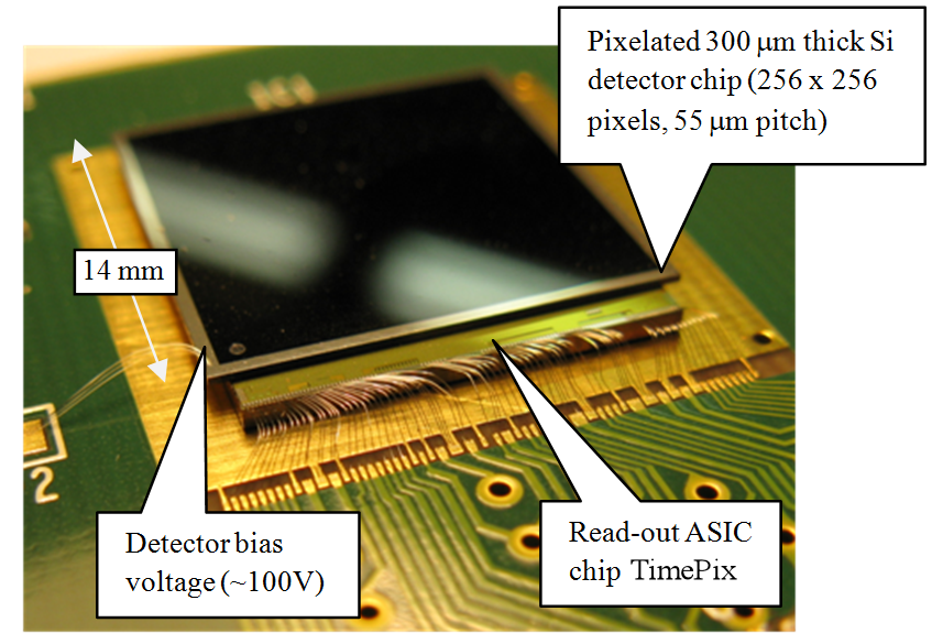
Each pixel is connected to its own readout chain. Timepix detector assemblies with different sensor materials (CdTe, GaAs and silicon) of different thicknesses (300 um to 1500 um) are available. Whereas the higher Z-materials are mainly used for X-ray imaging, Timepix detectors with silicon sensors have made their way into particle physics experiments, such as the ATLAS detector in the LHC, Space applications, and dosimetry in hadron therapy or in accelerator facilities.
Ionizing radiation creates free charge carriers (electrons and holes) in the sensor layer. These drift towards the corresponding electrode due to the applied electrical field, where they are finally collected. During their drift and collection, a current is induced at the electrodes of the pixel cells. The induced analog signal is shaped, amplified, and compared to a threshold level, which can be adjusted globally after a so-called threshold equalization. The voltage output signal is then used in different ways, depending on the selected mode:
- In counting mode, each time the analog output pulse exceeds the threshold, a counter in each pixel cell is incremented by one. Thus, the counter values correspond to the number of interactions in a given segment of the detector during the acquisition time.
- In Time-of-Arrival (ToA) mode, a counter is started once the signal crosses the threshold. It is stopped by the shutter signal terminating the data acquisition after the user defined exposure time.
- In Time-over-Threshold (ToT) mode, a counter is started once the signal crosses the threshold. It is stopped by the same signal crossing the threshold level again on its downward slope. The time interval the pulse stays above the threshold level can be calibrated to energy using fluorescence lines of known energy. Details on the calibration methods can be found here: http://aladdin.utef.cvut.cz/ofat/methods/TimePixCalibration/index.htm
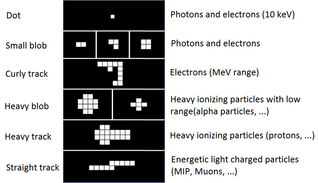
The detector segmentation allows for particle type differentiation by evaluating the characteristic imprints in the pixelscreens. A basic pattern recognition procedure can be used, e.g. to separate gammas, electrons or minimal ionizing particles (MIPs) from charged hadrons such as low energy protons, alphas or even heavier ions and neutrons. Figure 2 gives an overview of the basic cluster categories.
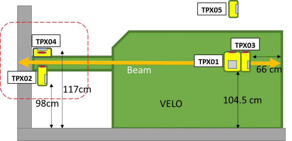
In the MoEDAL experiment 5 Timepix detectors with silicon sensor layers of thicknesses 300 um and 1 mm were installed. They are mainly used for the characterization of the radiation field at their positions, shown in Figure 3. However, due to their tracking capability, they are also able to identify highly ionizing events.